What is jaundice?
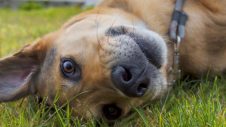
Jaundice is the accumulation of a yellow pigment, ‘bilirubin’, in the blood and in the tissues. It is most easily seen in the gums, whites of the eyes, and ear flaps. However, if these tissues normally have a dark colour, jaundice may not be seen.
What causes jaundice?
The causes of jaundice fall into three major categories:
1. Liver disease
Any disease that causes destruction of liver cells or causes bile to become trapped in the liver can cause jaundice.
2. Obstruction of the bile duct
The bile ducts carry bile (fluid essential for digestion) from the gall bladder to the small intestine. Obstruction can occur within the gall bladder or anywhere along the bile duct.
3. Destruction of red blood cells
Rapid destruction of red blood cells within the body leads to the production of bilirubin at a rate too high for the body to process and excrete it. The process of red cell destruction is known as ‘haemolysis’. It can occur within blood vessels (intravascular) or in the spleen and liver (extravascular).
How is the cause diagnosed?
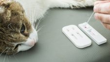
Within each category, there are several possible causes. Determining the cause of jaundice requires a series of tests. Some of these tests determine which category is involved. Once that is known, other tests are done to look for a specific disease which is leading to the jaundiced state.
What tests determine haemolysis?
Severe red cell destruction needs to occur before jaundice develops. An estimation of the numbers of red cells present can be done quickly and easily. Determination of red blood cell numbers is one of the first tests performed on the jaundiced patient.
What causes haemolysis?
Several tests are needed to determine which of the following is the cause:
- toxic plants
- chemicals
- drugs
- parasites on the red blood cells
- heartworm
- immune-mediated diseases
- cancer
What tests determine the presence of liver disease?
A biochemistry profile is performed on dogs with jaundice. This is a group of 20-30 tests that are performed on a blood sample. This profile contains several tests that are specific for liver disease. The main ones are the alanine aminotransferase (ALT), aspartate aminotransferase (AST), alkaline phosphatase (ALP), and the proportions of different types of bilirubin present in the blood.
Although each of these tests looks at the liver from a slightly different perspective, ultimately they only determine that liver disease is occurring. None of them is able to determine the exact cause of the disease. To make that determination, a biopsy of the liver is necessary. This can be done in three ways:
1. fine-needle aspirate
To perform this procedure, a small gauge needle is inserted through the skin into the liver. A syringe is used to aspirate some cells from the liver. The cells are placed on a glass slide, stained, and studied under a microscope. This is the least invasive and quickest test, but it has certain limitations. Because only a few cells are obtained, it is possible that a representative sample from the liver will not be obtained. Some diseases can be diagnosed with this technique and others cannot.
2. Surgical wedge biopsy
The dog is placed under general anaesthesia, and the abdomen is opened surgically. This permits direct visualisation of the liver so the exact site for biopsy can be chosen. A piece of the liver is surgically removed using a scalpel. This approach gives the most reliable biopsy sample, but carries the greatest risk since general anaesthetic and major surgery is involved.
3. Ultrasound guided biopsy
A large bore biopsy needle is used to obtain a small core of tissue, which is preserved in formalin and submitted to a pathologist. The area of liver being biopsied can be viewed and chosen using the ultrasound image as a guide. This technique carries fewer risks than a surgical wedge biopsy. Usually, a very short acting anaesthetic is necessary
What causes liver disease?
The most common causes of liver disease include:
- bacterial infections
- viral infections
- toxic plants
- chemicals
- drugs
- cancer
- immune-mediated diseases
- certain breed-specific liver diseases
What tests determine bile duct obstruction?
Dogs with obstructed bile ducts are usually extremely jaundiced. Their yellow colour can often be seen readily in the skin, as well as the whites of the eyes and the gums. However, an evaluation of the gall bladder and bile duct is necessary to be sure that obstruction is present.
An ultrasound examination is the most accurate non-invasive way to evaluate the gall bladder and bile duct. This technology uses sound waves to look at the liver, gall bladder and bile duct. If this is not available, X-rays should be taken of the liver. Sometimes exploratory surgery is necessary to properly evaluate the dog for biliary obstruction.
What causes bile dict obstruction?
The most common causes of bile duct obstruction include:
- pancreatitis
- trauma
- cancer
- gall bladder stones
- severely thickened bile
How is jaundice treated?
Jaundice is not a disease but rather a sign that disease is present. Therefore, there is not a specific treatment for jaundice. Jaundice will resolve when the disease that causes itself is cured. The basis for resolving jaundice is to diagnose the underlying disease. When the proper testing is done, this is usually possible. Then, treatment can begin.
If you have noticed discolouration in your pet’s ears, eyes, or mouth, or any other symptoms are present, contact your local Greencross Vets.